

We discuss many different eye conditions in our blog posts, because it’s important to us to raise awareness surrounding ocular issues that can affect anyone or their family. It’s also important for our patients to understand why we do certain things in an eye exam, and why we may request additional tests. Our patients’ eye health is our top priority, and we hope that by providing enough information, we can make that goal clear.
So, onward with today’s post.
You may have heard of Age-Related Macular Degeneration (also known as AMD). It usually comes up on the list of eye conditions that begin to affect people in the second half of their life, along with cataracts and glaucoma. AMD affects the central vision, creating blurriness and difficulty discerning details such as faces, driving conditions and reading.
Aptly named, it’s the most common cause of vision loss among folks over the age of 50, and is estimated to affect around 2.5 million Canadians. (When other factors outside of age are the culprit, the disease can manifest in younger people. In these cases, we drop the “A” and the diagnosis becomes simply Macular Degeneration.)
Below, we discuss the different types and stages of AMD, its causes, symptoms and treatments.

What Causes AMD?
At the back of the eye there is a small portion of the retina known as the macula. This little dude is responsible for our central and detailed vision. It contains a high-concentration of photoreceptor cells that send an electrical signal to our brain so it knows what we’re looking at.
Age is the biggest cause of AMD. As we age, our macula gets thinner. This makes it more susceptible to potential damage. Damage to the macula can result in blurriness, distortion or “wavy” vision, or dark areas, but all will be central; peripheral vision remains normal.
Other causes can include smoking (never good for the eyes), environmental factors, poor diet (especially diets high in saturated fats), high blood pressure, family history and genetics, and also race. AMD is most commonly found amongst Caucasian patients.
Dry vs. Wet
There are two main types of AMD: dry and wet.
Dry AMD is the most common. It occurs when the macula thins (hence, macular degeneration) and develops further when fat deposits known as drusen begin to form in the back of the eye, damaging the now fragile macula. Dry AMD is a progressive condition, meaning it will slowly worsen over time. The 3 stages are known as early, intermediate and late. Where someone falls on the scale depends on how far their condition has progressed.
Wet AMD (also known as neovascular AMD) is less common but considered more severe than dry AMD. It occurs when abnormal blood vessels begin to grow beneath the thinning macula. If these are not caught early, they can begin leaking blood and fluid into the eye, damaging the layer beneath the retina’s photoreceptors (the little dudes responsible for telling our brain what we’re looking at) and potentially scarring the macula. Untreated, wet AMD can cause severe loss of central and detailed vision, a loss that occurs much faster than with dry AMD.
Dry AMD can become wet AMD at any time, so it’s important to maintain regular exams with an optometrist (or ophthalmologist) to monitor all AMD symptoms. Regular exams and testing allow an eye doctor to catch the signs of AMD before the patient notices any vision loss, an invaluable asset to protecting your vision.
Geographic Atrophy
Geographic atrophy (or GA) is an advanced form of dry AMD caused when retinal cells begin to die. These areas of atrophy usually begin near the macula, causing distortion and blind spots in the central vision the macula is responsible for.
There doesn’t appear to be a clear direct cause of GA, but it’s believed to be linked to a combination of AMD factors, such as age, environment and genetics. Studies have found that GA in one eye makes it more likely to develop in the other as well.

AMD Symptoms
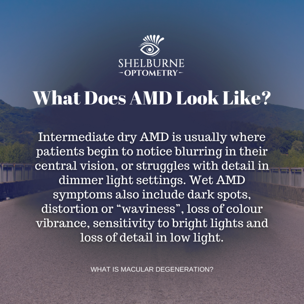
Early AMD symptoms are subtle, and not usually detected by patients. Intermediate dry AMD is usually where patients begin to notice blurring in their central vision, or struggles with detail in dimmer light settings.
Due to the increased speed of vision loss, wet AMD symptoms can seem to come on quite suddenly. They include blurred central vision, dark spots, distortion or “waviness” (think of a hot road on a summer day), loss of colour vibrance, sensitivity to bright lights and loss of detail in low light.
The symptoms are similar for GA, and at all stages of AMD the symptoms are painless, making early detection without the assistance of an optometrist or ophthalmologist impossible.
Detection
The best way to detect early stages of AMD are with a regular comprehensive eye examination with your optometrist. They will use drops to dilate your pupils, increasing the window needed to accurately assess the back of your eyes (where all this fun takes place).
It’s very likely they will recommend Optical Coherence Tomography (OCT Imaging) of the back of the eye, which takes photos and scans to create a clear picture of what’s going on with the surface and subsequent layers of the retina (including the macula).
With the help of the dilating drops and OCT, an optometrist can detect the early growth of drusen (those pesky fat deposits in the retina we mentioned earlier) and advise on how to slow the progression of the macula’s pending degeneration.
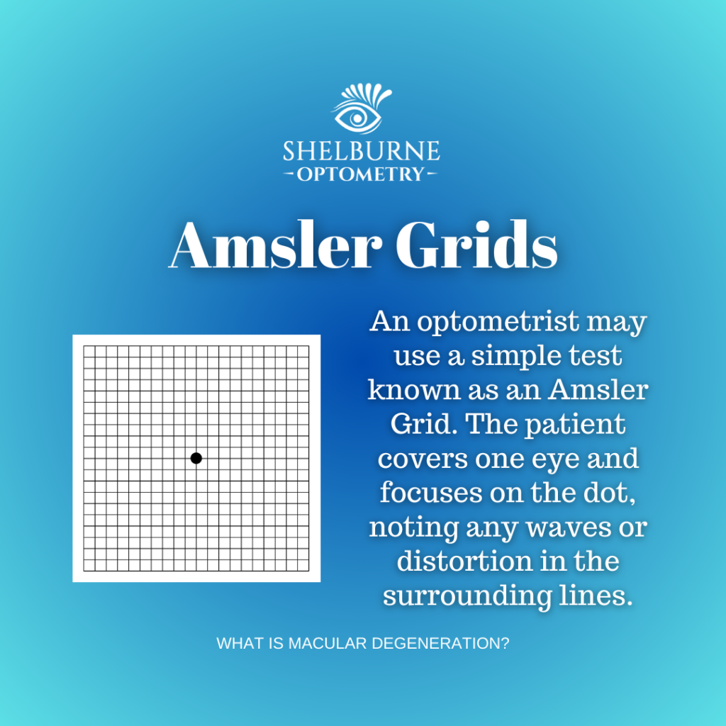
The optometrist may also employ the use of a simple test known as an Amsler Grid. This involves a paper showing a grid of little squares (similar to graph paper) with a dot in the centre. The patient covers one eye and focuses on the dot, noting any waviness or distortion in the surrounding lines of the grid. This is repeated with the other eye. A patient may also be sent home with a small pad of Amsler Grids to continue periodical testing at home, and contact their optometrist if they begin to notice any changes.
How Do We Treat AMD?
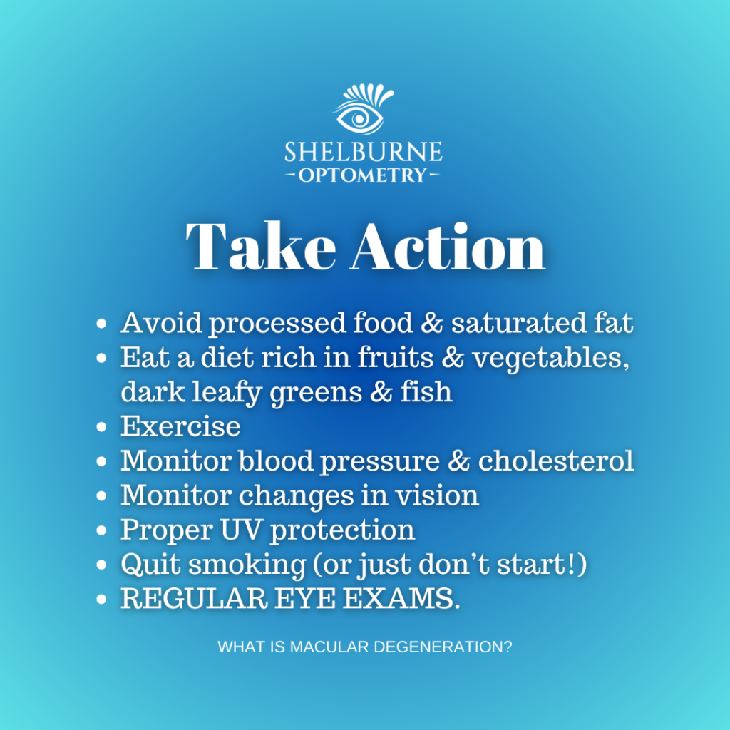
There are not currently any treatments for dry AMD, but there are ways to reduce its progression:
- Avoiding processed foods and anything high in saturated fats.
- Eating a diet rich in colourful fruits and vegetables, dark leafy greens, and fish such as mackerel, salmon and tuna to increase your body’s supply of healthy vitamins that benefit your ocular health.
- Exercise – healthy amounts of movement do wonders.
- Monitoring blood pressure and cholesterol levels.
- Monitoring changes in vision (both at home and with your regular eye exams).
- Proper UV protection (such as polarized sunglasses).
- Quitting smoking (or not starting to begin with – the ocular risks go beyond the back of the eye, including damage to blood vessels, tear and oil ducts, and the cornea).
- Regular eye exams.
In some cases, additional vitamin and mineral supplements may be recommended, such as:
- Copper
- Lutein
- Vitamins C & E
- Zeaxanthin
- Zinc
(Always consult your family doctor, eye doctor or ophthalmologist before starting any new supplements.)
Wet AMD does have treatment options, designed to slow the disease’s progression and prevent further vision loss. There are 3 types of treatment for wet AMD, and which one (or combination) is recommended is case-by-case:
- Anti-VEGF Treatments (injections) – Vascular endothelial growth factor (VEGF) occurs in the body and encourages formation of new blood vessels. Wet AMD increases VEGF, leading to an overgrowth of blood vessels in the eye. Anti-VEGF treatments are injected into the eye, blocking VEGF from working and preventing further vision loss (in some cases it has even been reported to reverse existing loss). There are various types of anti-VEGF drugs, and all work differently, have different side effects, and may have a different injection schedule. They’re usually the first treatment option for wet AMD.
- Photodynamic Therapy (PDT) – This treatment is performed by injecting a drug called verteporfin into the patient’s arm, which then travels to the blood vessels in the eye. The drug is then activated by a laser, reducing the leaking from blood vessels. It’s sometimes used in conjunction with anti-VEGF treatments, depending on the stage of wet AMD.
- Laser Surgery – When no other treatments are having the desired effect, a doctor may recommend laser surgery to seal abnormal blood vessels so they stop leaking into the back of the eye. It may slow down vision loss, but there is a risk of scarring and blind spots, so it’s generally viewed as a last resort.
There is currently no treatment for GA, but research is continually ongoing. Clinical trials are being done that include drugs, cell replacement therapy, gene therapy, and more, including retinal prostheses, to develop more treatments and options for patients living with AMD.
Final Thoughts
Macular degeneration is a common eye disease affecting millions of Canadians. While treatments are limited, early detection is possible and research is advancing all the time. Due to its focus on central vision, peripheral vision remains normal, and macular degeneration rarely leads to blindness.
The bottom line here is that regular comprehensive eye exams with your optometrist (preferably annually, if not bi-annually) are your best option for prevention and detection of these quiet eye diseases. If you’re one of those people who skip eye exams or additional testing because your “eyes are fine”, please consider the invaluable benefits of early detection, and the risks that increase when your ocular health isn’t a priority.
As always, we’re aiming to help you develop clear, beautiful memories to look back on, and prevent your risks of losing some of them to early asymptomatic stages of eye disease.
Because it’s a struggle to see, look and feel spectacular when you suddenly find yourself squinting all the time.
Wishing you all the best in wherever your path takes you today,
Sydney Gallant, CCOA