It’s World Glaucoma Week so we’re bringing awareness to this mysterious disease, and what it means to live with it in today’s world.
What is Glaucoma?
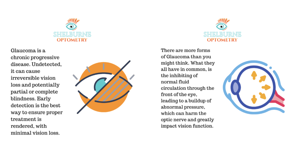
Glaucoma is a chronic progressive disease affecting the optic nerve, due to disruptions in fluid flow that increase pressure within the eye. Undetected, this disease can slowly eat away at your vision, causing irreversible vision loss and potentially partial or complete blindness. It is unbelievably important to be screened for glaucoma at your regular optometrist visits, especially if you have a history of the disease within your family. Early detection is the best way to ensure proper treatment is rendered, with minimal vision loss.
Forms of Glaucoma
There are more forms of glaucoma than you may think, and we’ve broken down the basics of each below. While each form has its own characteristics, the main thing they all have in common is the inhibiting of normal fluid circulation through the front of the eye, leading to a buildup of abnormal pressure inside the eye, which can harm the optic nerve and greatly impact vision function.

Glaucoma is a progressive disease occurring due to blocks in fluid flow.
Primary Open-Angle Glaucoma
Also known as chronic glaucoma, primary open-angle glaucoma is the most commonly diagnosed form.
Your eye is constantly producing its own fluid, known as aqueous fluid, which regulates eye pressure and carries essential nutrients to the eye’s tissues. It’s produced behind the iris (the pretty, coloured part of your eye), passes into the front of your eye through the pupil (yes—it really is a window), seeps through a sponge-like tissue called the trabecular meshwork, which releases it into the canal of Schlemm, which finally releases the fluid into the bloodstream.
If your eye begins producing too much fluid, or for some reason the fluid isn’t being drained properly, this puts pressure on the optic nerve, which results in higher pressure throughout the eye. The optic nerve is responsible for telling the brain everything your eye sees. When its fibres and blood vessels are under compression, they are especially susceptible to damage. This damage decreases the quality of information your eye sends to the brain, resulting in vision loss, which can be permanent if left untreated.
Primary open-angle glaucoma occurs when the trabecular meshwork (that spongy tissue the fluid needs to get through) gradually clogs or becomes blocked. This stops the fluid from being able to drain adequately, resulting in raised eye pressure. The cause of the clogging is unknown, but a common theory is that the eye’s drainage system may lose efficiency over time.
This form of glaucoma develops slowly and subtly, with no early symptoms to warn you. Generally, there is no pain or unusual cues until the peripheral vision starts to narrow, beginning to form a sort of tunnel vision. When this stage is reached, the peripheral vision loss may be permanent, but prompt treatment can preserve the remaining central vision.
Acute Glaucoma
Also known as angle-closure glaucoma, closed-angle glaucoma, or narrow angle glaucoma, acute glaucoma is not as common. While it can progress gradually, it is considered a medical emergency if it appears suddenly, as it can result in vision loss within a single day.
Acute glaucoma occurs when the angle between the iris (the pretty, coloured part) and the cornea (the protective tissue at the front of the eye) narrows or becomes blocked. This causes fluid buildup behind the iris, inhibiting its ability to drain. This results in immediate rise in eye pressure, which can lead to pain, severe headaches, nausea, and visual distortions. Treatment must occur within a few hours to avoid irreparable damage to vision. If treatment is successful, the vision is usually saved and another occurrence of acute glaucoma is highly unlikely.
Secondary Glaucoma
Secondary glaucoma occurs when the eye’s pressure is raised by factors outside its normal function. These can include a variety of medical conditions and abnormalities, medications, injuries, infections, or tumours present in or around the eye. This form of glaucoma can be temporary and curable if the cause is identified and removed in a timely manner. Secondary glaucoma is an excellent reason to report any past or recent eye injuries to your optometrist as soon as possible (to say nothing of possible retinal detachment, but we’ll talk about that in another post).
Normal-Tension Glaucoma
Also known as low-tension glaucoma, normal-tension glaucoma is more of a mystery. The eye pressure remains within a normal range, however, the optic nerve still becomes damaged. The cause for this remains unknown. There are recent studies that suggest it may be caused when the arteries supplying blood to the optic nerves begin to harden.
Congenital Glaucoma
Congenital glaucoma occurs when babies are born with defective drainage or circulatory systems in their eyes. Their vision is saved by opening the blocked passages surgically, to allow the fluid to flow more naturally.
Symptoms of Glaucoma
Glaucoma is a sneaky disease, and rarely shows early signs. There is often no pain or redness associated with its onset, and sadly, most undiagnosed patients realise something is wrong when their peripheral vision begins to darken. In most cases it’s too late to save this peripheral vision, but central vision can absolutely be saved with proper treatment and care.

Examples of how undetected glaucoma can manifest in your vision.
Who is at Risk
Glaucoma is not contagious or life threatening, and while it is very rare in young people, degenerative changes to the eyes as we age makes those over the age of 40 more vulnerable, and even more so those over 60. It is also more common in those with a family history of glaucoma (as it is considered hereditary to a certain degree), diabetics, those who are very near-sighted, and those who have sustained eye injuries or trauma. There are other factors within various medical conditions and eye anatomy that can lead to the onset of glaucoma.
How We Detect and Treat Glaucoma
The best way to detect glaucoma is with a comprehensive eye examination with your optometrist. In addition to checking the pressure of the eye (either with numbing drops and a tonometer, or with that super fun air puff test), your optometrist will also inspect the drainage angle inside the eye, as well as the general health of your retina and optic nerve. Optical Coherence Tomography (or OCT Imaging) may be requested for a closer look at the layers of the retina and optic nerve, and a Visual Field Test is an excellent way to check the range of your peripheral vision.
Glaucoma is treated through prescribed medications, usually in the form of drops or pills, and in some cases, surgery may be recommended to give the remaining vision its best chance. It takes time to adjust to the medication schedule, but regular and conscientious treatment can preserve a patient’s vision and allow them to continue living their lives fully, without concern of their condition worsening.
Final Thoughts
Your optometrist is always available to discuss any concerns you may have around your vision and eye health. No question is silly, and it will never bother your optometrist that you’ve chosen to take your eye health seriously. The internet is a wonderful source of information, but your eye doctor is still your best resource when it comes to your eye health.
At Shelburne Optometry we take glaucoma screening very seriously, and always do our best to have our patients leave our clinic confident that they’re getting the best care we can offer.
It is estimated that over 400,000 Canadians suffer from glaucoma and that around half of them aren’t even aware they have it. Prioritise your eye health, have your eyes tested regularly, and preserve your vision so you can continue making memories you’ll be grateful to look back on.
Wishing you all the best in wherever your path takes you today.


